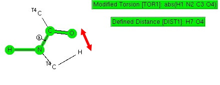
Amides with an H-N group are a component of the peptide linkage (O=C-NH). Here I ask what the conformation (it could also be called a configuration) about the C-N bond is. A search of the following type can be defined:
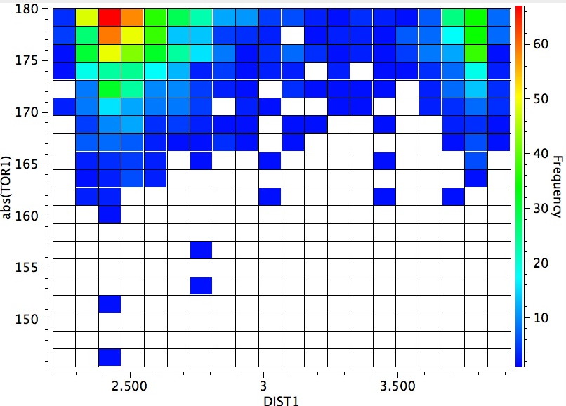
The dihedral shown is for H-N-C=O (but this is equivalent to the C-C-N-C dihedral, which is also often called the dihedral angle associated with the peptide group). I have also added a distance, from a C-H to the carbonyl oxygen. Other search constraints include T ≤ 175K, R < 0.05, no disorder, no errors, that neither N-C bonds are part of a ring and that the two carbons marked T4 both have four connected bonds. The search results in 619 hits (January 2013 version of the CCDC database), and these are displayed below.
The horizontal axis reveals the highest concentration (red) at ~2.4Å due to a syn-co-planar alignment of the C-H bond with the plane of the C=O bond in the s-cis conformer (the significantly smaller hot-spot at ~3.9A may be due to an anti-co-planar alignment of this C-H bond).
The vertical axis shows a clear preference for a dihedral of 179° (in fact no hits with a dihedral of less than 14o° were found) and this can only arise from the s-cis conformation in which the H-N bond is oriented antiperiplanar to the axis of the C=O bond. This preference can be rationalised by filled/empty NBO-orbital interactions, which include:
H-N/C=O. Click for 3D
Click for 3D.
This latter overlap conspires to bring the C-H hydrogen close to the oxygen (~2.35Å, DIST1 in the diagram above). So one might be entitled to ask: is this a hydrogen bond? There are (at least) two ways of testing this.
NCI surface. Click for 3D.
I end by reminding that the s-cis H-N-C=O conformation is a very common feature in peptides (the CCDC database comprises mostly small molecules, not larger peptides and proteins) arising from really quite subtle orbital interactions.
This post has been cross-posted in PDF format at Authorea.
In the mid to late 1990s as the Web developed, it was becoming more obvious…
I have written a few times about the so-called "anomeric effect", which relates to stereoelectronic…
The recent release of the DataCite Data Citation corpus, which has the stated aim of…
Following on from my template exploration of the Wilkinson hydrogenation catalyst, I now repeat this…
In the late 1980s, as I recollected here the equipment needed for real time molecular…
On 24th January 1984, the Macintosh computer was released, as all the media are informing…
View Comments